Born from the needs of a researcher
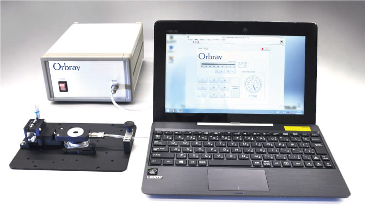
Product overview
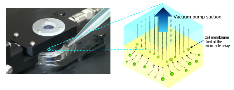
This product was born from the needs of a researcher who wanted to fix and observe cell membranes by using Φ2μm micro-hole array in silica glass. (This development is a collaboration with Associate Professor Takashi Okuno, Faculty of Science, Yamagata University.) By fixing cell membranes on a micro-hole array via suction, quantitative analysis of membrane protein can be done on an extremely tiny sample with an optical microscope.
The following video shows the actual images of HeLa cell membranes using the observation device and a fluorescence microscope.
Reference:Provided by Associate Professor Takashi Okuno, Faculty of Science, Yamagata University
Operation principles
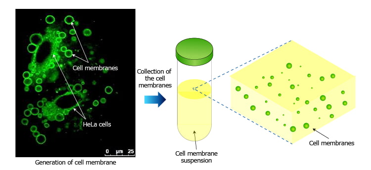
First, target sample cells are treated with a chemical solution and cell membranes are generated. Then the cell membrane suspension is prepared. (Please see the reference for protocol details.)
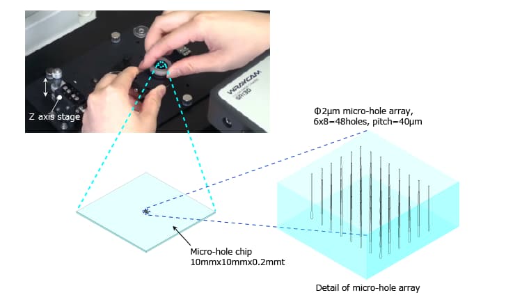
Micro-hole chip is set in the holder. The holder is connected to a vacuum pump via suction tube. Height of the chip (distance between the chip and a dish) can be adjusted by a z axis stage.
Cell membrane suspension is pipetted into the dish.
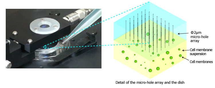
Desired pressure is selected on the computer, and suction started with the vacuum pump. The cell membranes are fixed at the micro-hole array.
Application examples
In this video, Φ6μm styrene beads are used to demonstrate cell alignment.
Provided by Associate Professor Takashi Okuno, Faculty of Science, Yamagata University
There are also other various applications in bio-research. This video shows fixing and observation of Tetrahymena.
Provided by Associate Professor Kentaro Nakano, Graduate School of Life and Environmental Sciences, University of Tsukuba
We have demonstration machines available to test the cell membrane observation device, so please feel free to contact us. Micro-hole designs are also customizable.
Specifications
| Specifications | ||
|---|---|---|
| Micro-hole chip | Hole diameter | 0.5μm∼20μm |
| Hole length | ≥ 0.2mm | |
| Aspect ratio | 100 | |
| Hole shape | Straight, tapered, hemispherical recess (see femtosecond laser) | |
| Material | Silica glass | |
| Standard chip |
|
|
| Customized chip | Hole diameter, layout, etc. can be customized. Please feel free to contact us. | |
| Chip holder for microscope | Transparent window | For phase-contrast microscope observation |
| Glass base dish | Compatible with Φ35mm diameter dish | |
| Z axis stage | For height adjustment of the micro-hole chip | |
| Suitable microscope | Olympus, Nikon (for other makes, please contact us) | |
| Outer dimensions | 170mm x 130mm x 58mmH | |
| Weight | 0.3kg | |
| Suction pump | Suction pressure | -1kPa∼-40kPa |
| Pressure adjustment resolution | 1kPa | |
| Maximum flow rate | 2L/min | |
| Control software | Pressure adjustment slide, pressure selection button, pressure increment/decrement | |
| Control PC | Laptop computer (not necessary if you have a computer. One USB port is needed.) | |
| Outer dimensions | 247mm x 160mm x 95mm | |
| Weight | 1.8kg | |
| Power source | AC100V∼250V | |
| Applications | Cell membrane observation, cell manipulation | |

